Signal Transduction During Animal Development
Defects in craniofacial development account for approximately a third of all human congenital birth defects. Communication between cells is important during embryonic development and defects in these signaling pathways are frequently also associated with disease in adults. Thus, understanding the causes of craniofacial birth defects and progress in developing therapeutics for adult disease are both hindered by our incomplete understanding of how cells normally process signaling information.
The success of the vertebrate lineage owes partly to the acquisition of jaws. The structures of the jaw apparatus also directly relate to function. Jaw development begins as cranial neural crest cells migrate from the neural tube into transient embryonic structures called the pharyngeal pouches. The neural crest cells surrounded by the first pharyngeal pouch form the chondrocytes that become the upper and lower embryonic jaw cartilages. The adult craniofacial skeleton forms later through elaborating upon and largely replacing these embryonic jaw cartilages.
The Endothelin-1 (Edn1) signaling pathway is required for lower jaw formation. The Edn1 signal emanates from the pharyngeal pouches to instruct the neural crest cells. First, the Edn1 ligand binds to a G Protein-Coupled Receptor (GPCR) on the neural crest cells that couples to a G-alpha-q factor to communicate with a Phospholipase C (PLC) family member. Next, PLC stimulation leads to the activation of a group of transcription factors that further modulate the expression of a downstream network of genes. Hence, we understand the series of events set in motion by the Edn1 signal fairly well. However, induction of edn1 gene expression is not well understood.
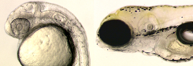
Wdr68 is required for edn1 expression and is capable of physically interacting with two members of the Dual-specificity tyrosine-regulated kinase (Dyrk) family. Strikingly, wdr68 mutants lack both the upper and lower jaws. Thus, our current focus is on elucidating the roles that two factors, Wdr68 and Dyrk1b, play in animal development. Through a combination of genomic and developmental methods using zebrafish mutants and antisense gene knockdown models, we are identifying and characterizing the genes and pathways essential for normal craniofacial development. We hope these efforts will lead to a better understanding of embryonic development and new routes for therapeutic intervention.
